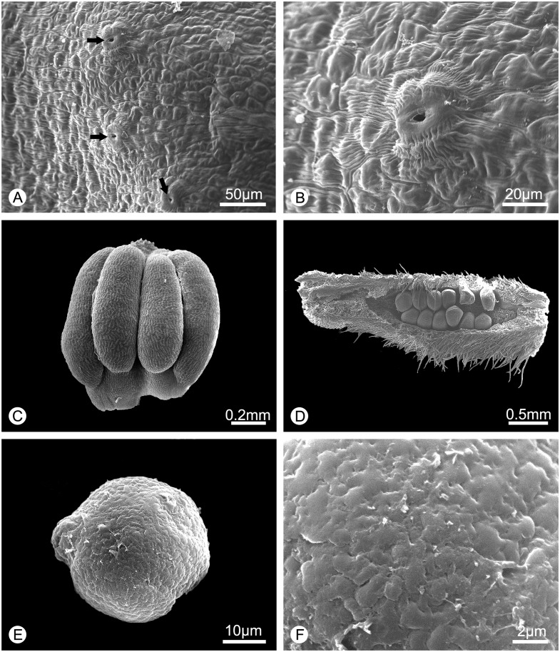
Fig 3. Morphology of the surface of the inner petal glands, stamen, carpel and pollen of Alphonsea glandulosa (scanning electron micrographs).
A, Surface of the glandular tissue, showing the nectary stomata (arrowed). B, Close-up of the nectary stomata. C, Stamen. D, Carpel, dissected to show biseriate ovules. E, Pollen grain. F, Rugulate pollen exine ornamentation.

