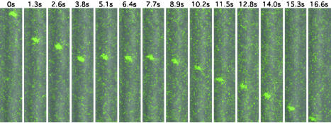
Fig. 1.
Retrograde transport of GFP-capsids after entry. Shown is fluorescent time-lapse recording of an axon of a DRG sensory neuron. Images were captured by laser-scanning confocal microscopy of the field every 0.638 s after viral inoculation onto the cells (every other frame is shown). Differential interference contrast images were simultaneously captured by a back-side detector and were overlayed with the fluorescence images. A single capsid is seen moving from the top of the field to the bottom. The pause in motion is typical of the overall saltatory motion. The axon of the infected neuron was oriented with the cell body below the field of view, consistent with retrograde motion. (Area of field shown = 5.6 × 28.0 μm.) For a time-lapse recording of entry dynamics, see Movie 1, which is published as supporting information on the PNAS web site.

