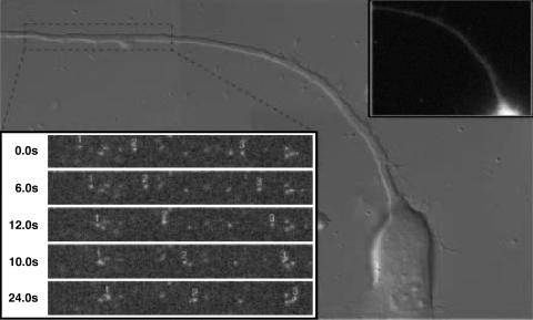
Fig. 4.
Entry transport is not blocked in a late-stage infected neuron. In superinfected neurons, newly entering viruses display net retrograde motion typical of normal infections. Neurons were first infected with PRV-GS791, which expresses a GFP-capsid but is deleted for the gD glycoprotein. The deletion inhibits the herpesvirus block to superinfection, allowing for subsequent infection with a second virus (PRV-GS847, which expresses a mRFP1-capsid). Superinfection was begun at 13.5 h post initial infection, allowing for PRV-GS791 progeny virus to egress before addition of PRV-GS847. The imaged neuron is shown in the background in three overlapping differential interference contrast images. GFP imaging showed the neuron to be in the late stages of infection from the first virus (PRV-GS791), as indicated by the oversaturated fluorescence emitting from the soma (due to capsid assembly and maturation) and two capsids in the axon (Inset, top right). Retrograde capsid transport of PRV-GS847 was monitored by red emission, and three capsids undergoing entry transport toward the soma are shown (Inset, bottom left; area of field shown = 68 × 9 μm). Additional dim punctae are extracellular virions that bound to the axon shaft but did not enter (and were partly bleached during imaging).

