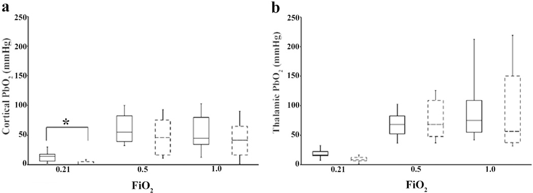
Figure 2.
Cortical (a) and thalamic (b) PbO2 for sham surgery (solid boxes) and CA animals (dashed boxes), at 24 h after CA while the rats were administered different oxygen concentrations: 50% oxygen (FIO2=0.5), 100% oxygen (FiO2=1), and room air (FiO2=0.21). The box plots display the median ± one standard deviation, and maximum and minimum values. Cortical and thalamic hypoxia is present when the rats inhale FiO2=0.21. (*p<0.016 vs. sham) (n=6–8/group)

