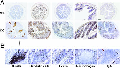
Fig. 2.
Characterization of leukocyte infiltrates in the inflamed Runx3 KO colon. IHC on WT and KO colon sections (×4) (A) and on a KO B cell cluster (×10) (B) with anti-CD45R for detection of B cells, anti-IgA for detection of IgA, anti-GFP for detection of DC (13), anti-CD3 for detection of T cells, and anti-F4/80 for detection of macrophages. (Scale bars: A, 250 μm; B, 50 μm.)

