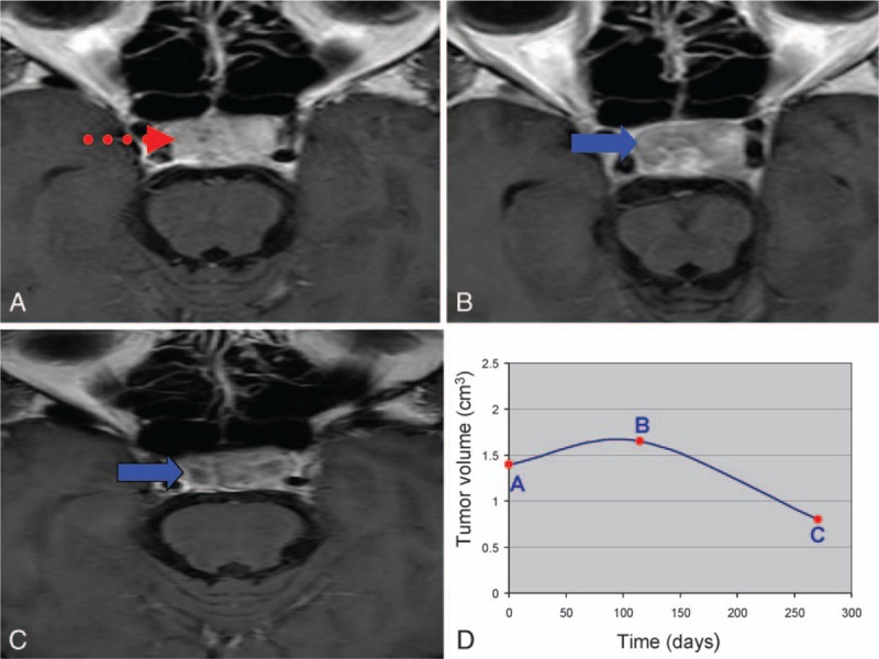
Figure 12.

(A) Baseline magnetic resonance imaging (MRI) of pituitary adenomas before CyberKnife stereotactic radiosurgery (CK SRS). The dotted arrow indicates the contrast enhanced area. (B) Follow-up MRI at the 4th month post-CK SRS demonstrated transient progression and reduced flow of gadolinium contrast (solid arrow). (C) Follow-up MRI at the 10th month post-CK SRS demonstrated volume regression and reduced flow of gadolinium contrast (solid arrow). (D) The curve of volumetric change corresponded to different time points A, B, and C.
