Figure 3.

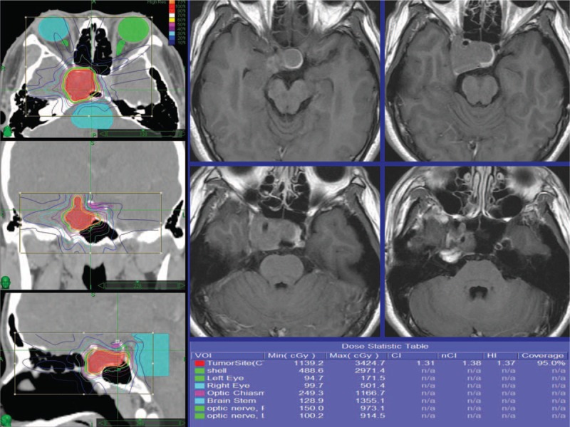
Radiosurgery dose plan images demonstrating a pituitary adenoma which was eventually controlled. The radiosurgery dose planning is shown with contours of the tumor and brainstem and optic chiasm.

Radiosurgery dose plan images demonstrating a pituitary adenoma which was eventually controlled. The radiosurgery dose planning is shown with contours of the tumor and brainstem and optic chiasm.