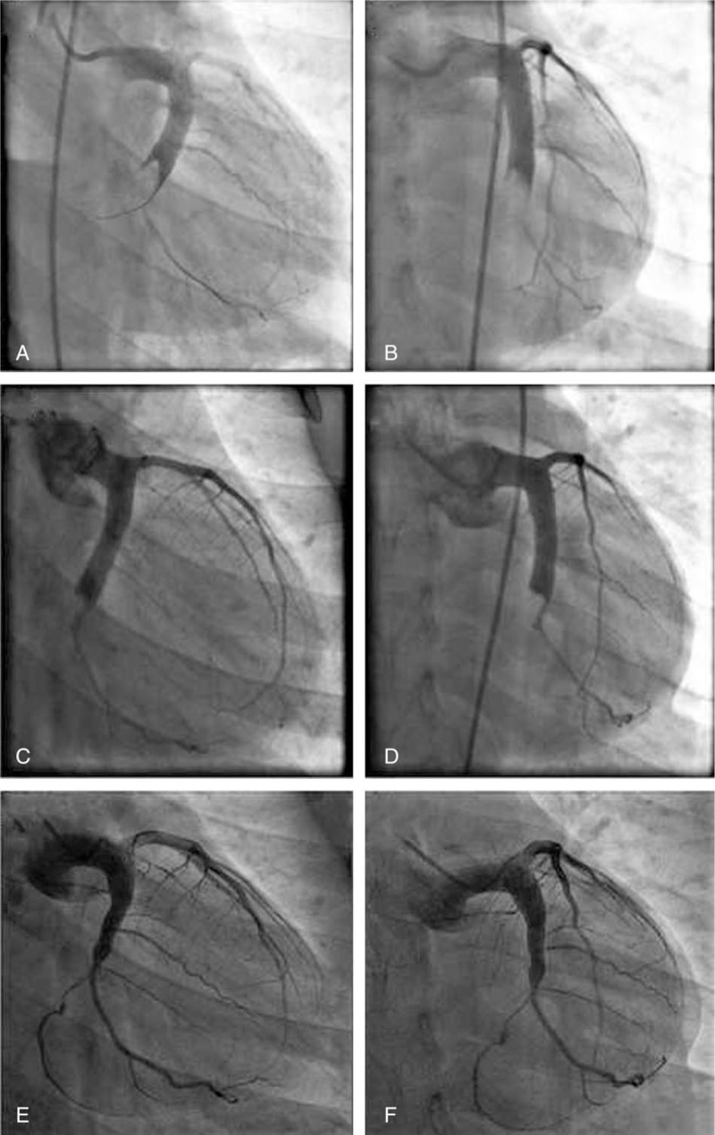
Figure 2.

Angiographic images of the LCX, postthrombus aspirations in (A) right caudal view and (B) antero-posterior caudal view. After several attempted thrombus aspirations, only TIMI 2 flow was restored in the second obtuse marginal branch. The second coronary angiography images of the LCX 15 days after admission in (C) right caudal view and (D) antero-posterior caudal view. There was still an obstructive filling defect in the distal portion of the LCX. TIMI 3 flow was restored in the second obtuse marginal branch. The third coronary angiography images of the LCX 15 months after discharge in (E) right caudal view and (F) antero-posterior caudal view. TIMI 3 flow was restored in the LCX and almost complete resolution of the thrombus was noted. LCX = left circumflex artery, TIMI = thrombolysis in myocardial infarction grade.
