
Figure 3.
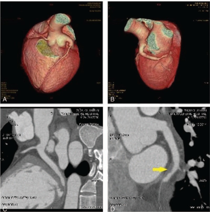
Sixty-four-slice coronary computed tomographic angiography images of the LCX 10 days after admission. A and B, Three-dimensional computed tomography reconstruction showing coronary artery ectasia involving the LM and LCX. C and D, A filling defect is shown in the distal portion of the LCX (arrow). LCX = left circumflex artery, LM = left main artery.