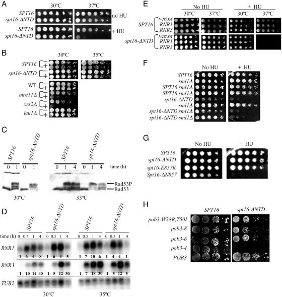
Figure 7.
The Spt16 NTD mediates HU sensitivity. (A) Five-fold serial dilutions of isogenic spt16Δ derivatives maintained by a plasmid-borne spt16-ΔNTD or SPT16 gene were spotted onto rich medium and incubated at the indicated temperature in the presence or absence of 50 mM HU. (B) Growing cells were transferred to rich medium with (+) or without (−) 200 mM HU, incubated for 8 h, and then serially diluted, transferred to HU-free solid medium, and incubated at the indicated temperature for colony formation. Isogenic mre11Δ and xrs2Δ cells (67) were positive controls for sensitivity, while isogenic leu1Δ and wild-type (WT) cells were negative controls. (C) Rad53 protein was detected by western analysis (using antibody from Santa Cruz Biotechnology) in extracts of growing cells incubated for the indicated times in rich medium containing 200 mM HU. Rad53P indicates phosphorylated Rad53. (D) RNR1 and RNR3 RNAs in extracts of growing cells incubated for the indicated times in rich medium containing 200 mM HU were detected by northern analysis, and quantified (bolded values in each lane) relative to TUB2 RNA values by densitometric analysis using multiple exposures and ImageJ software (US National Institutes of Health; available at http://rsb.info.nih.gov/nih-image/). (E) Serial dilutions of stationary-phase cultures of cells harbouring the RNR1 and RNR3 expression plasmids pBAD070 and pBAD079, or the control plasmid pBAD054, were spotted on rich medium with or without 50 mM HU and incubated at the indicated temperature. (F) Cells with the sml1Δ deletion (67) and isogenic derivatives with tagged SPT16 or spt16-ΔNTD at the chromosomal locus were spotted on rich medium with or without 50 mM HU and incubated at 30°C. Two segregants of sml1Δ derivatives are shown. (G) Five-fold serial dilutions of isogenic spt16Δ derivatives carrying the indicated plasmid-borne spt16 allele were spotted onto rich medium with or without 50 mM HU and incubated at 30°C. (H) Serial dilutions of cultures of the strains shown in Figure 6D were spotted on rich medium containing 100 mM HU and incubated at 30°C.

