
Figure 4.
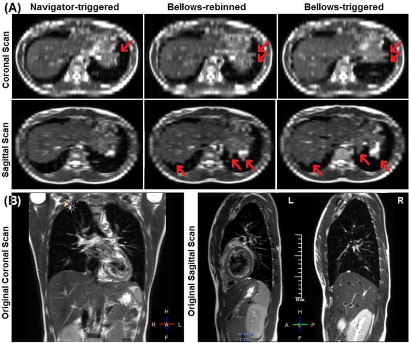
Illustration of binning artifacts of navigator-triggered, bellows-rebinned (concurrent), and bellows-triggered (consecutive) 4DMRI images (volunteer 6) (A). The axial views are re-sliced from the coronal or sagittal scan images (B). The right diaphragm dome appears smooth in navigator-triggered 4DMRI, while both sets of bellow-based 4DMRI contain similar motion artifacts with irregular edges (red arrows).