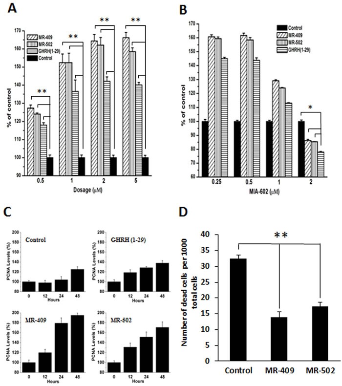
Figure 2. Stimulation of proliferation and inhibition of apoptosis of human dermal fibroblasts by GHRH agonists.

A. Primary human dermal fibroblasts were treated by 0.5-5 μM GHRH agonist or GHRH(1-29) in serum-free Fibrolife medium. The numbers of living cells at day 4 were chemiluminescently quantified. Error bars represent SEM, **p < 0.01. B. Cell proliferation in the presence of 1 μM GHRH agonists and 0.25-2 μM MIA-602, a GHRH antagonist. Error bars represent SEM, *p < 0.05. C. Fibroblasts were treated by 2 μM of GHRH (1-29), MR-409, or MR-502 in serum-free medium for 48 hours. The proliferating cell nuclear antigen (PCNA) expression levels were measured by western blots. Error bars represent SEM. D. Cell viability assay was conducted under the conditions of serum depletion. Living and dead cells in minimal 20 random fields were counted. The numbers of dead cells in a total of 1000 cells were calculated and shown in the plot. Error bars represent SEM, **p < 0.01.
