
Figure 6. Acceleration of healing of skin wounds by topical application of GHRH agonist, MR-409.
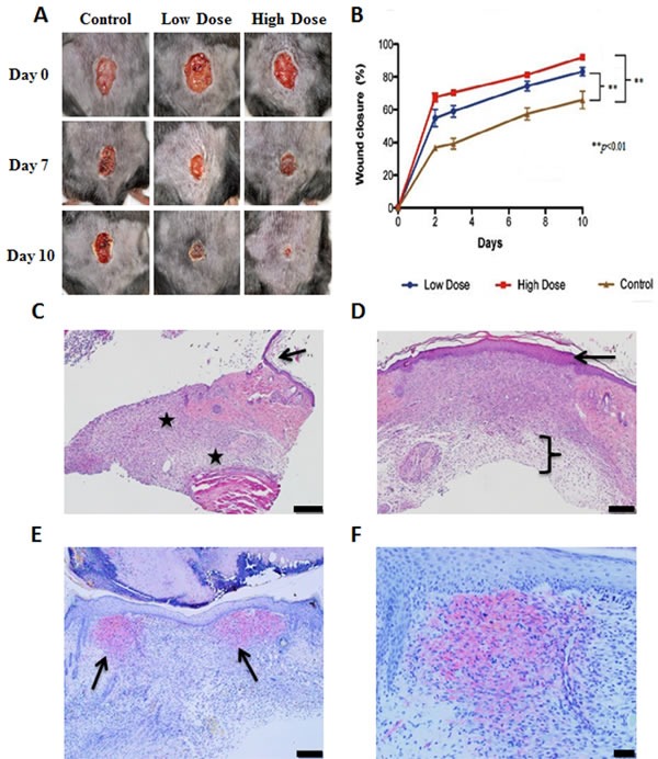
A.. Representative photographs of the wounds at 0, 7 and 10 days after treatment with 1 μg/day or 10 μg/day MR-409, or vehicle. The pictures were taken from the same animals at selected days. B. Morphometric analysis. Photographs were taken on days 0, 2, 3, 7 and 10 and analyzed to determine the percentage of wound closure. Error bars represent SEM, **p < 0.01. C. Photomicrographs of control wounds at day 10, H&E stained. Black stars indicate extensive fibrosis and scarring. The arrow points to the edge of the wound. Due to the extensive fibrosis, the epidermis is thin and poorly attached to the dermis. Scale bar represents 0.2 mm. D. Photomicrographs of treated group, H&E stained. The bracket indicates less fibrosis in the dermis vs control. The epidermis is thicker and appears better adhered to the dermis (arrow). Scale bar represents 0.2 mm. E. Treated wound, stained for αSMA. Arrows indicate two areas of positive staining at the edge of the wound. Scale bar represents 0.2 mm. F. Treated wound, stained for αSMA. Higher magnification. Scale bar represents 0.05 mm.