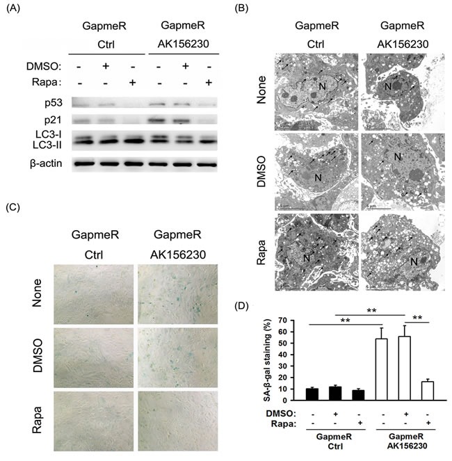
Figure 4. Rapamycin enhances autophagosome formation and rescues cellular senescence induced by AK156230 knockdown in MEFs.

A. Western blotting for LC3, p53 and p21 using lysates from the indicated cells. β-Actin was used as the loading control. B. Transmission electron microscopy images of MEFs treated with 2.5μM rapamycin or DMSO for an additional 48h after transfection with control GapmeR control or GapmeR AK156230 for 24h. Black arrowheads indicate representative autophagosomes or autophagolysosomes, and the nucleus is denoted by N. These sections were examined at 120kV with a JEOL JEM-1400 transmission electron microscope. C. Representative images of the SA-β-gal activity staining for the indicated cells. D. Percentage of the indicated cells positive for SA-β-gal activity was shown. Cells were quantitated by randomly choosing at least four independent fields. Student's t-test, **P < 0.01.
