
Figure 2. The levels of IKKα and p65 phosphorylation during skin aging.
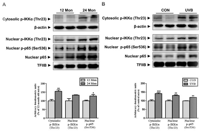
Western blotting was performed to detect the levels of IKKα phosphorylated at Thr23 and p65 at Ser536 in skin homogenates from 12- and 24-month-old mice (A) and in skin homogenates of control and UVB-irradiated mice (B) Blots were quantified by densitometry and normalized to β-actin or TFIIB. Bars represent the mean percentage value ± SEM in 12-month-old mice (n = 7-8, * p < 0.05, ** p < 0.01 vs. 12-month-old mice) and control mice (n = 7-8, * p < 0.05, ** p < 0.01, *** p < 0.001 vs. control mice).