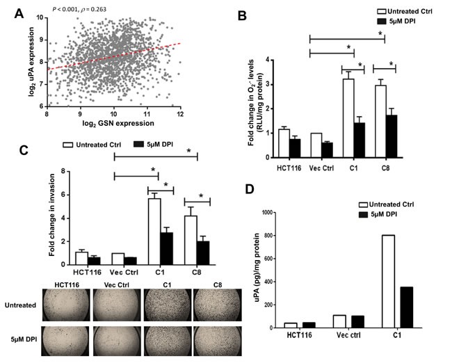
Figure 5. DPI inhibits gelsolin-induced O2.- invasion and uPA levels.

(A) Correlation of gelsolin expression with uPA across 1820 colorectal cancer samples. Spearman's rank correlation test was used to access the correlation of uPA expression with gelsolin. The correlation coefficient (ρ) and its significance (P value) are indicated. (B-D) Treatment of cells with 5μM DPI for 24 hours significantly lowered (B) O2.- levels and (C) invasion of gelsolin-overexpresing cells and (D) gelsolin-induced uPA secretion. Upper panel B, quantitative representation of invaded cells following 5μM DPI treatment. Lower panel B, representative pictures of invaded cells with or without DPI treatments are shown (2.5X magnification of the entire well). *p-value <0.05 versus controls using a two tailed Student's t-test. Values (mean ± SD ) are expressed as fold over the empty vector control, which was arbitrarily set as one. (D) Cells were serum starved with or without 5μM DPI for 8 hours and the conditioned media were used to detect uPA by ELISA. Treatment of cells with 5μM DPI significantly inhibited uPA secretion in the gelsolin-overexpressing C1 cells whereas 5μM DPI treatment minimally affected uPA secretion in the empty vector control and wild-type HCT116. Secreted uPA levels were normalized to protein concentration. The data shown here is the raw ELISA reading and a representative of three independent experiments.
