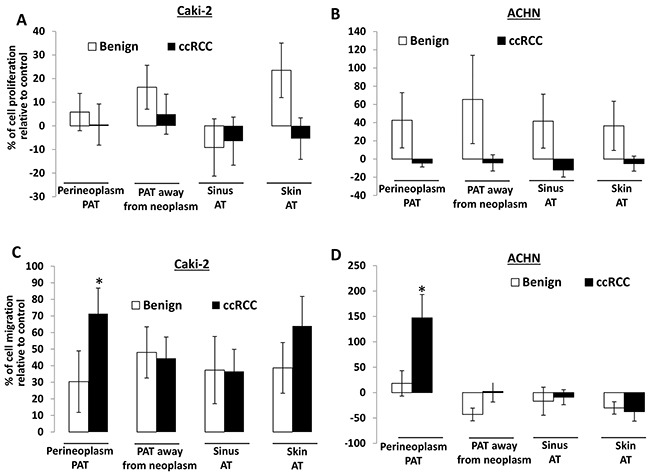
Figure 1. The effect of different AT CMs on proliferation and migration of Caki-2 and ACHN cells.

A. 2×10⁴ Caki-2 or ACHN cells were plated in 24-well culture plates. After 24 hours, the medium was changed to serum-free medium (negative control) or CMs from different AT cultures, including perineoplasm PAT, PAT away from neoplasm, renal sinus and skin ATs from patients with ccRCC or benign renal diseases. After 72 hours of incubation, cell densities were measured by MTT assay. Columns, mean percentages of cell proliferation under treatment of AT CM relative to negative control; bars, SEM. Experiments were replicated thrice. B. cells were applied to the upper surface of a membrane. After incubation for 48 hours, the upper surface of the membrane was scrubbed free of cells; the membrane was fixed, H&E stained, and photographed. Representative pictures were taken from the lower surface of three independent membranes at ×100 magnification. Number of migrated cells was counted from 10 random fields. Columns, mean percentages of migratory cells under treatment of different AT CM relative to negative control; bars, SEM. Experiments were replicated thrice. “*” denotes P < 0.05.
