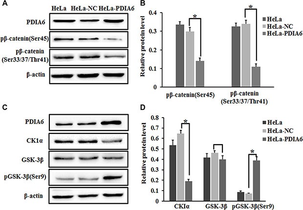
Figure 4. PDIA6 suppresses the phosphorylation of β-catenin.

(A) HeLa cells were transfected with pCMV-sport6-PDIA6 or pCMV-sport6 and collected to detect PDIA6, phosph-β-catenin Ser45 and phosph-β-catenin Ser33/Ser37/Thr41 by Western blot. PDIA6 overexpression resulted in a reduction in the amount of phosphorylated-β-catenin normalized to control cells. (B) The quantitative analysis of these proteins was normalized to β-actin. *p < 0.05. (C) Immunoblotting was performed to detect CK1α-GSK-3β and pGSK-3β (Ser9) levels after HeLa cells transfected for 48 h. (D) The quantitative analysis of these proteins was normalized to β-actin. *p < 0.05.
