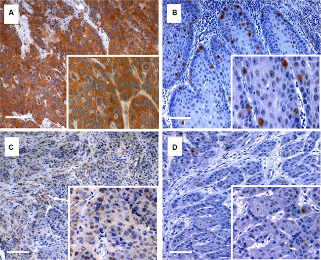
Figure 1. Immunohistochemical staining of Plk3 and pT273 caspase-8.

Examples of anal cancer biopsies with high (A, C) and low (B, D) immunohistochemical detection of Plk3 and pT273 caspase-8. Original magnification × 100 (inlets × 400), scale bar: 100 μm.
