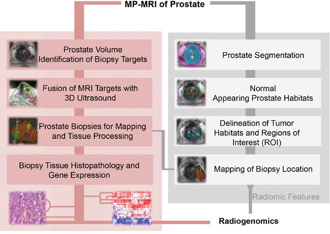
Figure 1. Experimental Design.

Multiparametric (mp)MRI, consisting of anatomical (T2-weighted), perfusion (Dynamic Contrast Enhanced [DCE]-MRI) and diffusion (Diffusion Weighted Imaging [DWI]) imaging sequences is acquired on 3T scanner. Upper left-hand side (shaded in pink) denotes the procedures for mpMRI-ultrasound fused targeted biopsies. The steps for radiomic analysis are presented at the right hand side in grey. Histopathology results, gene expression analysis and radiomic features are combined in the radiogenomic analysis.
