
Figure 5. Radiation-induced IL6 secretion mediates activation of Notch signaling.
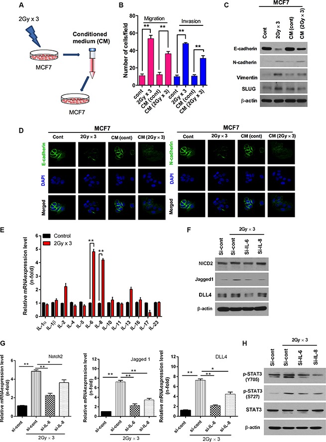
(A) Schematic model illustrating conditioned medium treated MCF7 breast cancer cells. MCF7 cells were irradiated 2 Gy × 3 as mentioned earlier. Then cell's medium was further harvested and treated to another MCF7 cells. (B) Migration and invasion assay in transwells after irradiated cultured medium those are compared with directly irradiated MCF7 cells. (C) Western blot and (D) immunocytochemistry for EMT markers after fractionated irradiation of MCF7 that are treated with irradiated cultured medium. (E) qRT PCR analysis for mRNA levels of cytokines family in MCF7 cells after fractionated irradiation. (F) Western blot for NICD2, Jagged1 and DLL4 after fractionated irradiation of MCF7 that are transfected with siRNA targeting IL-6 and IL-8. (G) qRT-PCR for Notch2, Jagged-1 and DLL4 mRNA levels after irradiation of MCF7 cells that are transfected with siRNA targeting IL-6 and IL-8. (H) Western blot for activation of STAT3 after fractionated irradiation of MCF7 that are transfected with siRNA targeting IL-6 and IL-8. β-actin was used as a loading control. Error bars represent mean ± S.D. of triplicate samples. *p < 0.05 and **p < 0.01.