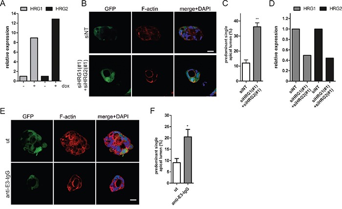
Figure 6. HRG depletion restores lumen formation in oncogenic K-RasG12V expressing Caco-2 cells.

A. Caco-2tet K-RasG12V cells were seeded into 3D in the presence of dox. Three days after seeding, cysts were isolated, RNA was extracted, and HRG1/HRG2 expression was determined by qPCR and normalized to GAPDH (N=2). (B-D) Caco-2tet K-RasG12V cells were transfected with non-targeting (siNT) and a mix of HRG1- and HRG2-specific siRNAs (#1). The next day cells were seeded into 3D culture and K-RasG12V expression was induced with dox one day later. B. Cultures were fixed three days later and stained with DAPI (nuclei; blue), anti-aPKC antibody (cyan) and phalloidin (F-actin; red). GFP is coexpressed with K-RasG12V (green). Shown are confocal sections of the midplane of representative cysts (scale: 20 μm). C. The percentage of cysts with PSAL from (B) was determined (n>70; N=3). D. Cysts were isolated three days later and RNA was extracted. HRG1/HRG2 expression was determined by qPCR and normalized to GAPDH (N=2). E. Cells were seeded into dox-containing 3D culture in the absence (ut) or presence of 100 nM anti-E3-IgG. Three days later CTX was added. Cultures were fixed the next day and stained with DAPI (nuclei; blue), anti-aPKC antibody (cyan) and phalloidin (F-actin; red). GFP is coexpressed with K-RasG12V (green). Shown are confocal sections of the midplane of representative cysts (scale: 20 μm). F. The percentage of cysts with PSAL from (E) was determined (n>70; N=3).
