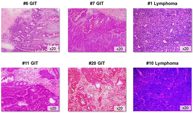
Figure 1. Tumor histology.

Representative H&E sections of GIT and lymphomas from MLH1-/- mice. GIT appeared as well-differentiated adenocarcinomas showing different invasive potential and morphology. Non-Hodgkin lymphomas were of either B- or T cell origin. Original magnification x20.
