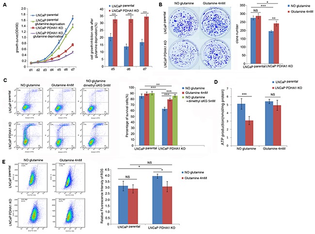
Figure 4. Effect of glutamine on cell growth, apoptosis, ATP and ROS production.

A. shows the growth inhibition due to glutamine deprivation was significantly higher for the PDHA1 KO cells compared to the parental cells, especially at day5 (P=0.001), day6 (P<0.001) and day7 (P<0.001). The bars on each detection point represent standard deviations from three independent experiments. B. shows the PDHA1 KO cells cultured in the medium with glutamine form significantly more clones (P=0.006) compared to the cells cultured without glutamine, while the parental cells cultured with glutamine shows no significant difference compared to the cells cultured without glutamine (P=0.760). C. shows strong glutamine addiction of the PDHA1 KO cells for survival. The PDHA1 KO cells with glutamine depletion exhibit significantly higher apoptosis rate compared to the cells with 4.0mM glutamine (P<0.001) and 5mM dimethyl supplement (P<0.001), while no significant apoptosis rate is observed between the parental cells cultured without glutamine and neither with glutamine (P=0.094) nor 5mM dimethyl αKG (P=0.059). D. Glutamine addition in the medium recovers the ATP production in the PDHA1 KO cells. When the cells were cultivated in the medium without glutamine, there are 5.1193 and 3.0758nmol ATP/mg protein in the parental and PDHA1 KO cells, respectively (P<0.001). However, when both of the cells were cultivated in the medium with glutamine, 5.4200 and 4.9408nmol ATP/mg protein could be observed (P=0.217) in the parental and PDHA1 KO cells, respectively. The measured ATP concentration was normalized by total protein concentration in each sample. The data are presented as means ± S.D (n=5). E. Glutamine addition in the medium decreased the ROS production in the PDHA1 KO cells. Left panel is the representative images of DCFH flow cytometry of each group. The right panel is the relative mean DCFH fluorescence intensities presented as percentage relative to the control value. There are significantly less ROS detected in the PDHA1 KO cells cultured in medium with glutamine compared to the cells cultured in the medium with glutamine depletion (P=0.016), while there is no significant difference of ROS detection for the parental cells cultured in medium with or without glutamine (P=0.443). The data are expressed as mean ± S.D. of 3 independent experiments. *P < 0.05, **P < 0.01, ***P<0.001, according to the 2-tailed Student's t test.
