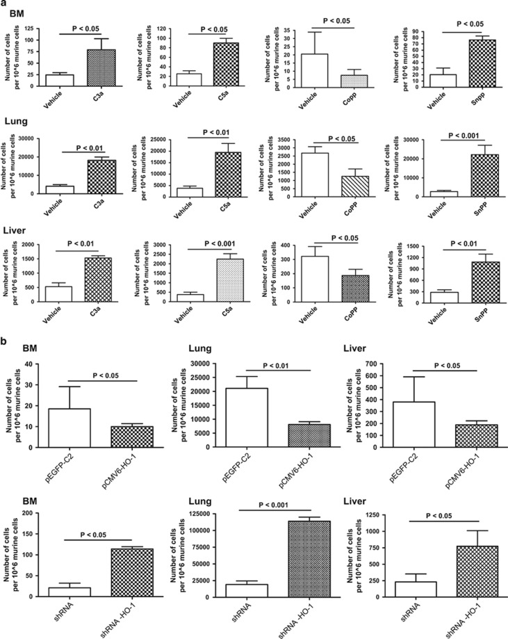
Figure 5.
Complement C3 and C5 cleavage fragments and downregulation or inhibition of HO-1 enhance metastasis, whereas upregulation or activation of HO-1 inhibits the metastasis of leukemic cells in vivo. (a) Detection of transplanted human U937 cells (10 × 105 per mouse) in the organs of irradiated (SCID)-beige inbred mice after in vivo transplantation. Pre-implantation, the cells were incubated ex vivo with vehicle only, C3a (1 μg/ml), or C5a (140 ng/ml) for 6 h and HO-1 activator (CoPP, 50 μmol/l) or HO-1 inhibitor (SnPP, 50 μmol/l) for 2 h. Under all conditions, serum-free medium was used. Human cells were detected in BM (top row), lung (middle row) and liver (bottom row) by the presence of human Alu sequences in purified genomic DNA samples according to RT-qPCR. (b) Evaluation of the spread of transplanted leukemic cells in vivo after up- and downregulation of HO-1. Human HO-1-overexpressing (RAJI-pCMV6-hHO-1) and knockdown (RAJI-shHO-1) hematopoietic cell lines and RAJI-pEGFP and RAJI-shRNA cells as control counterparts, respectively, were prepared for in vivo transplantation (10 × 105 per mouse). Twenty-four hours after cell transplantation into irradiated SCID mice the organs were collected, and detection and quantification of the human cells was then analyzed by RT-qPCR. For statistical comparisons, a one-way analysis of variance and a Tukey's test for post hoc analysis were carried out, and means±s.d. are shown. Significance levels are indicated at P<0.05, P<0.01 and P<0.001 for comparison with untreated or control-transfected cells.

