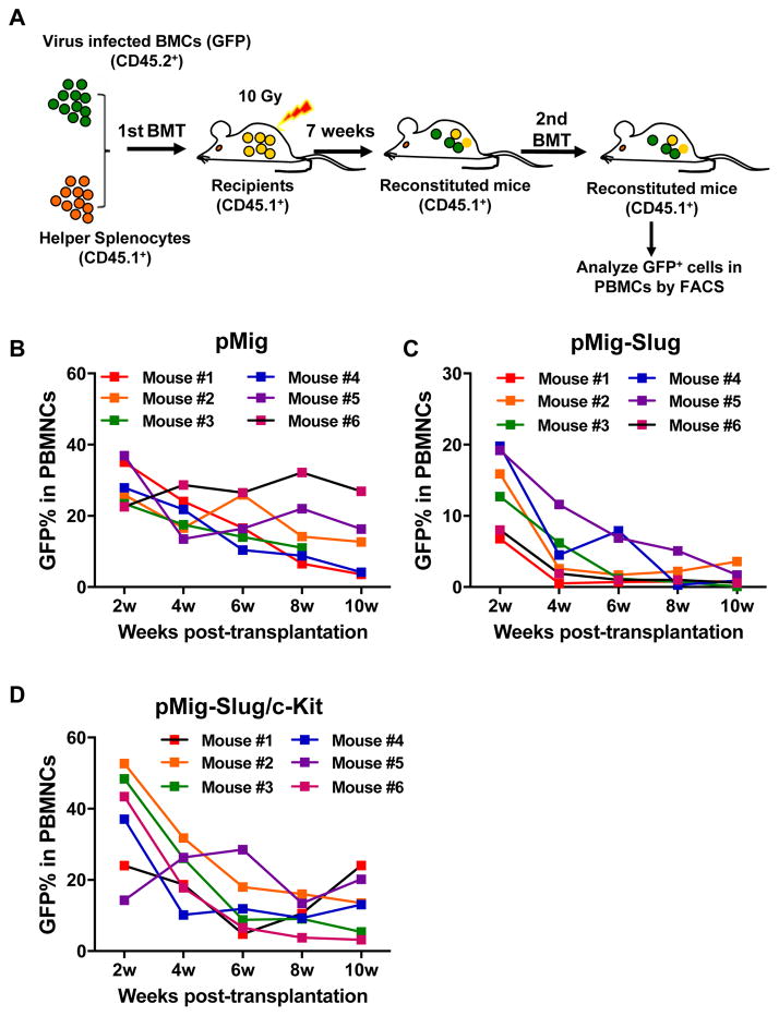
Figure 5. Serial BM Transplantation Assay for HSCs Exhibits the Fine regulatory Interaction between Slug and c-Kit.
(A) Diagram of serial BM transplantation assay for in vivo HSC. CD45.2+ BM cells were transduced with retroviral particles containing pMig vector, pMig-Slug, or pMig-Slug/c-Kit, and then transplanted with helper splenocytes (CD45.1+) into lethally irradiated mice (CD45.1+). By 7 week of transplantation, primary BM cells were isolated from reconstituted mice and performed second transplantation. The percentage of GFP+ cells in peripheral blood was assessed every two weeks after second transplantation.
(B–D) Analysis of GFP percentage in peripheral blood by flow cytometry in pMig- (B), pMig-Slug (C), or pMig-Slug/c-Kit (D) reconstituted mice (n = 6). Data are representative of two independent experiments.
See also Figure S9.

