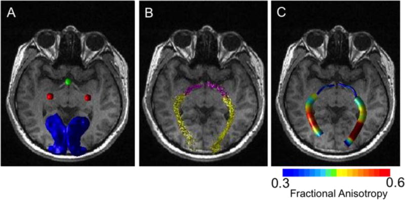
Figure 3.

(A) Typical ROI seeds used for fiber tracking overlaid on an anatomical image from a representative normal subject: optic chiasm3, lateral geniculate nucleus (red), and V1 (blue). (B) Fiber tracts of the optic tract (pink) and optic radiation (yellow). (C) The corresponding fractional anisotropy along the fiber tracts.
