Fig. 3.
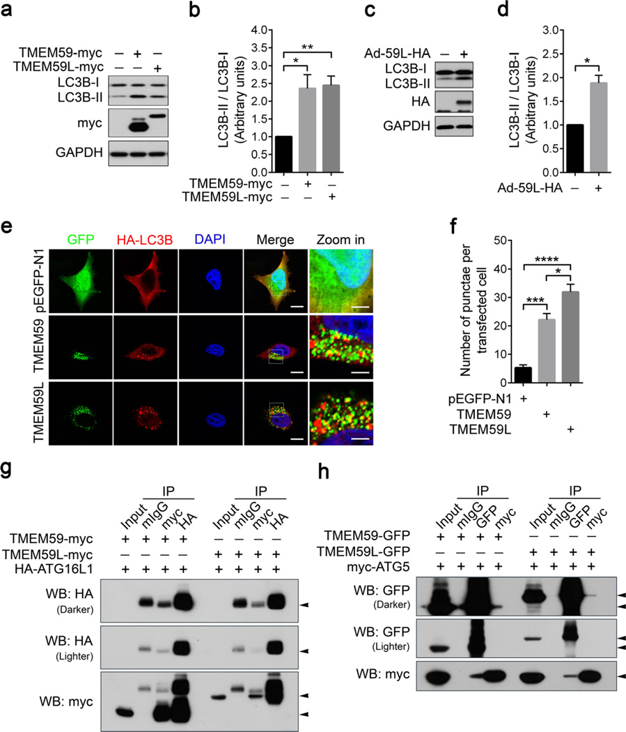
Both TMEM59L and TMEM59 interact with ATG5 and ATG16L1 and their overexpression induces autophagy. a, b HEK293T cells were transfected with TMEM59-myc, TMEM59L-myc, and control vector for 36 h. a Equal amounts of protein lysates were subjected to Western blot for LC3B-I/II. b Protein levels of LC3B-I and LC3B-II were quantified by using densitometry and normalized to those of GAPDH for comparison. n = 3, *p < 0.05, **p < 0.01. c, d Mouse primary cortical neurons were transduced with control adenoviruses (−) or adenoviruses expressing TMEM59L (Ad-59L-HA, +). Cells were harvested after 4 days. c Equal amounts of protein lysates were subjected to Western blot for LC3B-I/II. d Protein levels of LC3B-I and LC3B-II were quantified by using densitometry and normalized to those of GAPDH for comparison. n = 3, *p < 0.05. e, f Hela cells were co-transfected with TMEM59-GFP or TMEM59L-GFP and HA-LC3B for 24 h. e After immunostaining with anti-HA antibody and incubating with secondary antibody, cells were observed under a confocal microscope. Red indicates HA-LC3B, green indicates GFP, and blue indicates nuclei. Scale bars: 10 µm (“zoom in” images); 20 µm (all other images). f The number of HA-LC3B-positive vesicles per transfected cell was counted for at least ten cells. n = 3,*p < 0.05, ***p < 0.001, ****p < 0.0001. g Myc-tagged TMEM59 and TMEM59L were co-transfected with HA-tagged ATG16L1 into HEK293T cells. Equal amounts of cell lysates were subjected to immunoprecipitation (IP) with mouse normal IgG (mIgG), mouse antibody against myc, or mouse antibody against HA, followed by Western blot (WB) analysis. h GFP-tagged TMEM59 and TMEM59L were co-transfected with myc-tagged ATG5 into HEK293T cells. Equal amounts of cell lysates were subjected to IP with mIgG, mouse antibody against GFP, or mouse antibody against myc, followed by WB analysis. Five percent of cell lysates were used as input

