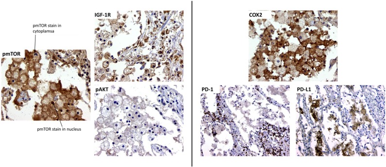
Figure 1.
Morphoproteomic analysis of human TB lung sample. Left: stain for phosphorylated mTOR, insulin-like growth factor-1 receptor (IGF-1R), and phosphorylated Akt at 400× magnification. Right: sample stained with anti-human cyclooxygenase 2 (COX-2) and visualized at 400×. Programed death-1 (PD-1) and programed death-1 ligand (PD-L1) stain, magnification at 200×.

