Figure 5.
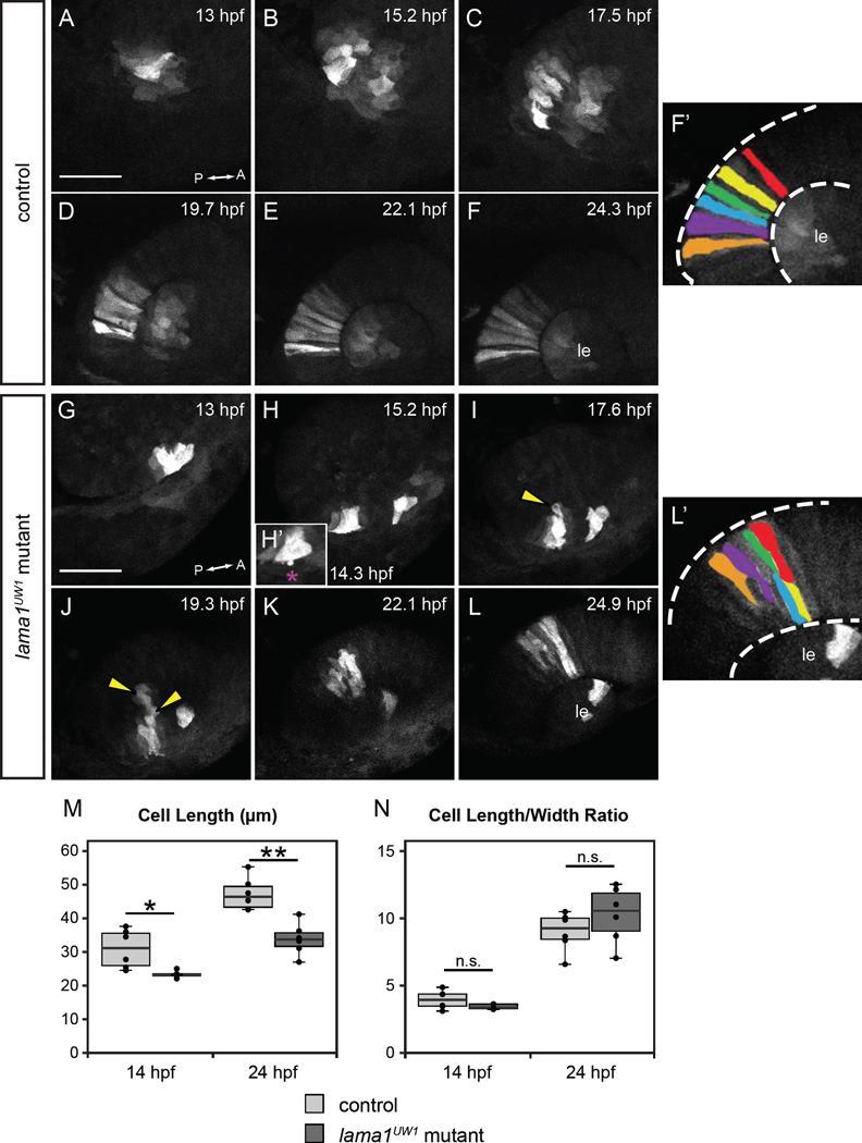
Loss of motile cell behaviors does not underlie optic cup morphogenesis defects in lama1UW1 mutant embryos.
Retinal progenitors expressing Kaede were exposed to 405 nm light, which converts Kaede from green to red fluorescence via an irreversible photocleavage. Images are maximum intensity projections from 4-dimensional datasets of the red (converted) channel.
(A-F’) Images from a timelapse of a lama1UW1 control sibling embryo. (G-L’) Images from a timelapse of a mutant embryo. (H’) Zoomed image of single timepoint (14.3 hpf) showing a retinal progenitor extending a bleb (magenta asterisk) beyond the boundary of the optic vesicle. (F’, L’) Pseudocolor of marked retinal cells. Retinal progenitors in the control embryo extend across the entire width of the retina, while retinal progenitors in the lama1UW1 mutant embryo elongate, but do not span the width of the retina. Loss of apicobasal register marked by arrowheads.
(M, N) Comparisons of length (M) or length/width ratio (N) of retinal progenitors in lama1UW1 mutant or control embryos. While retinal progenitors are longer in control embryos than mutants, the length/width ratio is not significantly different. *P<0.02; **P<0.001; n.s. = not significant
Dorsal views; scale bar, 50 µm. le, lens; dashed lines, retina margins. A, anterior; P, posterior.

