Figure 6.
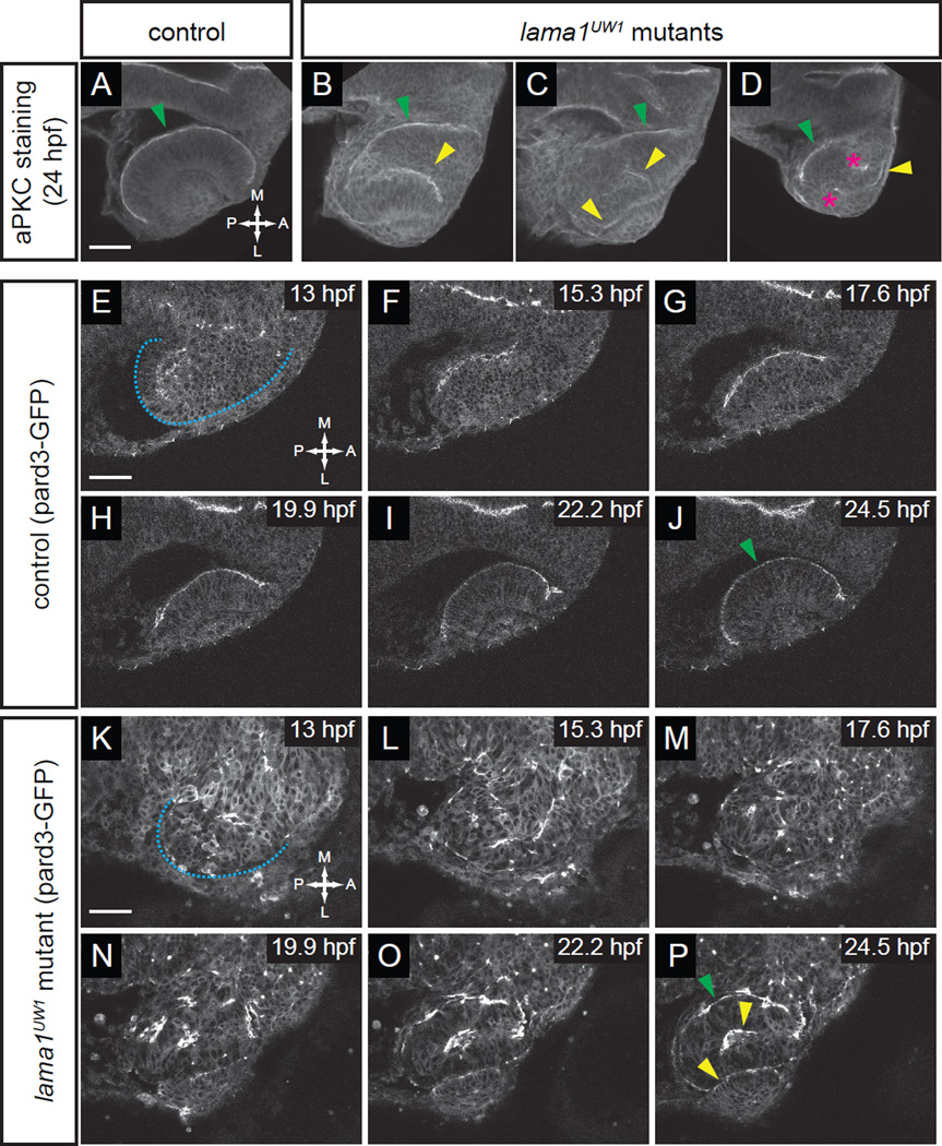
Apicobasal polarity is disrupted from the earliest stages of optic vesicle morphogenesis.
(A–D) Antibody staining for aPKC in control (A) and lama1UW1 mutant embryos (B–D) reveals disruption of polarity at 24 hpf. green arrowheads, correct apical domain. yellow arrowheads, ectopic apical domains. asterisks, ectopic puncta.
(E–P) Single confocal sections from 4D datasets of apical domain dynamics (marked by pard3-GFP) in a lama1UW1 control embryo (E–J), or a mutant embryo (K–P). In lama1UW1 mutant embryos, pard3-GFP localization is disrupted similar to aPKC. dashed blue line marks outline of optic vesicle. green arrowheads, correct apical domain. yellow arrowheads, ectopic apical domains.
Dorsal views; scale bar, 50 µm. A, anterior; P, posterior; M, medial; L, lateral.

