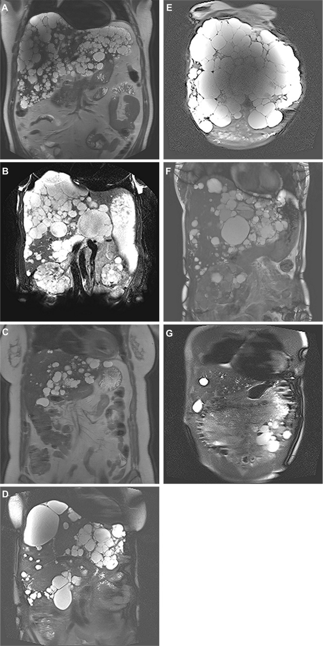
Fig. 1.
Baseline T2-weighted magnetic resonance imaging of patients developing hepatic cyst infection during the DIPAK-1 Study. Height-adjusted liver volume (hTLV) were 2670, 1579, 823, 2723, 8635, 1901, and 997 mL/m in respectively, cases 1–7 (a–f). In five of seven patients, the phenotype consists of multiple small- and medium-sized cysts spread throughout the liver, with remaining large areas of non-cystic liver parenchyma [cases 1–4 (a–d) and 6 (f)]. One patient [case 5 (e)] showed a phenotype with massive diffuse involvement of liver parenchyma by small- and medium-sized liver cysts, with only a few areas of remaining normal liver parenchyma between cysts. Remarkably, the liver phenotype of the last patient who developed a hepatic cyst infection was limited to a single hepatic cyst [case 7 (g)]

