
Figure 1.
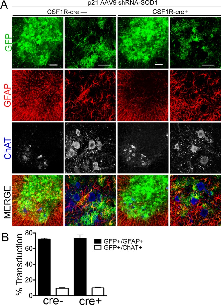
AAV9 transduces astrocytes when injected in adult CSF1R‐cre+ and cre− SOD1‐G93A mice. (A) Immunohistochemistry and quantification of lumbar spinal cords of SOD1‐G93A; IKK β F/wt; CSF1R‐cre− mice (left) and SOD1‐G93A; IKK β F/wt; CSF1R‐cre+ mice (right) injected at postnatal day 21 with AAV9‐SOD1‐shRNA expressing GFP. (B) Quantification of transduced (GFP+) cells at postnatal day 100 shows no significant difference in the mean percent of transduced astrocytes (GFP+/GFAP+) between CSF1R‐cre− (72.2 ± 0.7%) and CSF1R‐cre+ mice (73.2 ± 2.5%). Few motor neurons were transduced at p21 in both CSF1R‐cre− (9.6 ± 0.5%) and CSF1R‐cre+ mice (10.1 ± 0.6%), determined by counting transduced cells (GFP) co‐labeled ChAT+ motor neurons throughout the lumbar spinal cord. Lumbar spinal cords from three animals per group were analyzed. Scale bars indicate 100 μm (left) and 50 μm (right). AAV9, adeno‐associated viral vector serotype 9; GFP, green fluorescent protein.