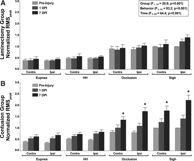
Fig. 5.
Bilateral chronic diaphragm EMG activity for animals in the laminectomy (A; n = 9–12) and C4 unilateral contusion (B; n = 12) groups. Summary of diaphragm RMS EMG amplitude (RMSpeak) at preinjury (light gray) and 1 (dark gray) and 7 (black) days postinjury (DPI) normalized to the preinjury RMS EMG amplitude for sigh. In the repeated-measures analysis, there was an effect of group, behavior, and time. Animals in the laminectomy group displayed stable chronic diaphragm EMG values for all respiratory motor behaviors across the 8-day recording period. At 7 DPI, animals in the contusion group displayed significantly higher ipsilateral and contralateral RMS EMG amplitudes compared with the preinjury and 1 DPI values during occlusion and sighs (*post hoc Tukey-Kramer HSD at P < 0.05). HH, hypoxia-hypercapnia.

