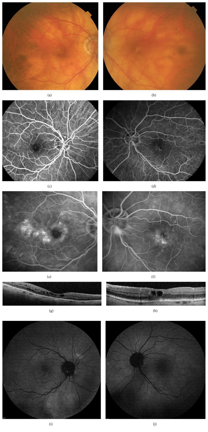
Figure 1.
Patient 1. (a), (c), (e), and (g): right eye. (b), (d), (f), and (h): left eye. (a) and (b): fundus examination showing small pigment epithelium alterations in the foveal area without obvious vascular abnormalities. (c), (d), (e), and (f): fluorescein angiography (min and min) showing perifoveal telangiectasia with late intraretinal staining. (g) and (h): spectral-domain optical coherence tomography (B-scan) showing cystoid macular edema with a central macular thickness measured to 312 μm on the right eye and to 345 μm on the left eye. (i) and (j): autofluorescence images were unremarkable.

