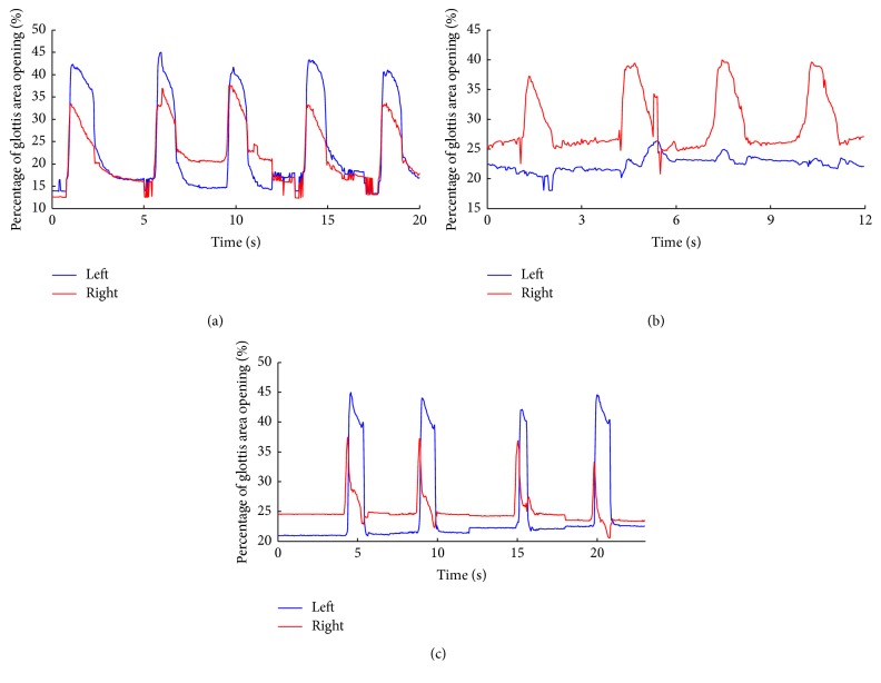
Figure 5.
Displacement of vocal fold across experiment conditions. The movements of left (blue) and right (red) vocal fold were separately calculated. (a) Changes of glottis area in the intact state. (b) Changes of glottis area following left RLN destruction. (c) Changes of glottis area following pacing with feedback from CT EMG. The left (injured) and right (healthy) sides of glottis movements were plotted using blue and red solid lines, respectively.

