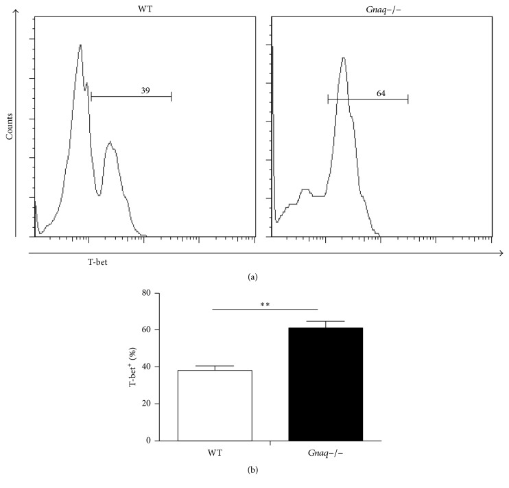
Figure 3.
Loss of Gαq enhances the expression of T-bet. (a) Purified naïve CD4+ T cells from WT and Gnaq−/− mice were stimulated with anti-CD3/CD28 (3 μg/mL), in the presence of mouse IL-12 (20 ng/mL), mouse IL-2 (20 ng/mL), and anti-IL-4 (10 μg/mL) for five days. Cells were harvested, fixed, permeabilized, and stained with PE-cy7-conjugated anti-T-bet and analyzed by flow cytometry. (b) The percentage of T-bet+ cells was calculated. All data are presented as mean ± SD; ∗∗P < 0.05, n = 3. The result is representative of three independent experiments.

