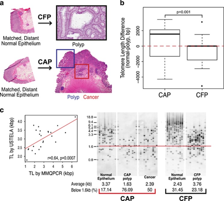
Figure 1.
Telomere length distinguishes cancer-adjacent polyps (CAPs) from cancer-free polyps (CFPs). (a) Corresponding tissue of CAP and CFP models that are used in this study. CFP cases include matched, distant normal colon epithelium, and the villous adenoma (polyp). All polyp cases used in the study were matched by histology and degree of dysplasia—villous adenomas with low-grade dysplasia. CFP cases are those that have had polyps present and removed that have not gone on to cancer. CAP cases include matched, distant normal colon epithelium, the polyp (residual polyp of origin) and the corresponding cancer that arose from the polyp. The cancer specimens from CAP cases represent a range of all stages. (b) Boxplots showing the values for the difference in telomere length in the polyp from the matched normal colon epithelium, for both CAP and CFP polyp cases from left to right, respectively. First, the telomere length for each polyp was subtracted from its matched normal telomere length to obtain a difference in telomere length value. Positive difference values indicate that the telomere length in the polyp tissue was shorter than the matched normal, and the inverse for the negative difference values (positive values, shorter polyp telomeres than normal; negative values, longer polyp telomeres than normal). Red, dashed line is drawn at zero for ease in determining positive and negative differences. The telomere length difference between CAP and CFP polyps is significant, P=0.001. (c) Scatterplot (far left) showing the correlation in telomere lengths in kilo base pairs (kbp) as determined from the modified monochrome quantitative PCR (MMQPCR) method and Universal STELA (USTELA) method, showing a strong, positive correlation. The other panels show results from USTELA from left to right, an exemplar CAP case and CFP case, showing normal, polyp, and cancer for the CAP and normal and polyp for the CFP. The horizontal red line indicates the threshold at which the load of short telomeres is determined (below 1.6 kb). In addition, the average telomere lengths in kilobases (kbs) and the percentage of telomeres below the 1.6 kb threshold (%) were both quantified and are included at the bottom of each electropherogram. There is an increasing amount of short telomeres from normal to polyp to cancer in the CAP case, with a slight telomere length increase in the cancer relative to the polyp (but still overall shorter compared with normal). Overall short telomeres occur to a lesser degree in the CFP polyp compared with normal as well as with the CAP tissues.

