Figure 10.

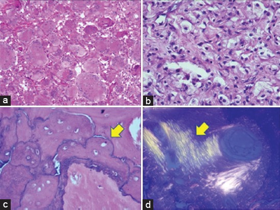
Histopathology. (a) Initial biopsy showing classical features of a giant cell tumor, including uniform sprinkling of osteoclast-like giant cells with intervening mononuclear stromal cells. Hematoxylin and eosin (H and E) staining ×200, (b) sections from post denosumab-treated specimen displaying replacement by foamy histiocytes and lymphocytes. H and E staining ×400, (c) section from host bone displaying “jigsaw puzzle”- like or mosaic pattern of lamellar bone, pathognomonic of Paget’s disease. One of the several thickened mosaic cement lines marked with arrowhead. H and E staining ×200, (d) disorganized collagen fibers arranged in several directions rather than uniform (arrowhead), seen under polarized light. H and E, ×200.
