Figure 2.

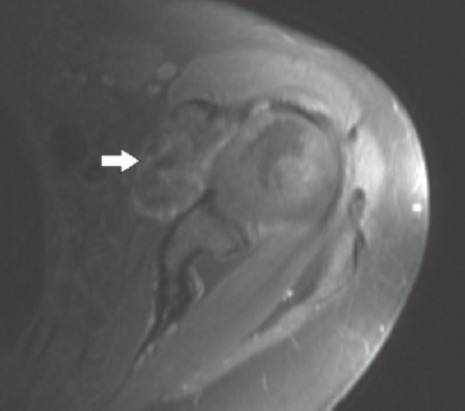
Axial magnetic resonance image after gadolinium contrast shows the mass to have peripheral and septal enhancement (white arrow).

Axial magnetic resonance image after gadolinium contrast shows the mass to have peripheral and septal enhancement (white arrow).