Figure 4.

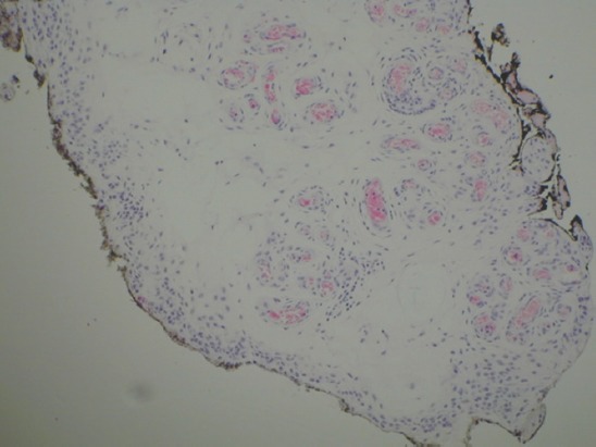
Synovium with thick-walled blood vessels and scattered lymphocytes, consistent with synovial hemangioma (Hematoxylin and Eosin, ×40).

Synovium with thick-walled blood vessels and scattered lymphocytes, consistent with synovial hemangioma (Hematoxylin and Eosin, ×40).