
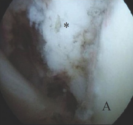
Figure 6.
Arthroscopic image of the mass (asterisk) extending through rotator interval just before open excision. Glenoid (A) is in the right lower corner.
Official websites use .gov
A
.gov website belongs to an official
government organization in the United States.
Secure .gov websites use HTTPS
A lock (
) or https:// means you've safely
connected to the .gov website. Share sensitive
information only on official, secure websites.
Arthroscopic image of the mass (asterisk) extending through rotator interval just before open excision. Glenoid (A) is in the right lower corner.