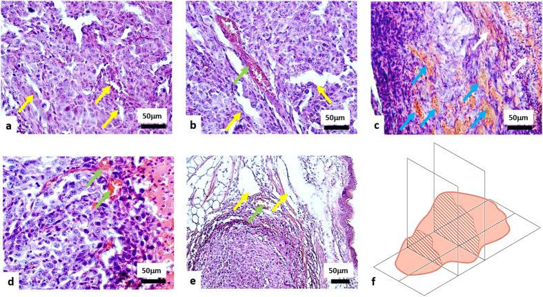
Figure 2. H&E histology of CT26 tumours 7 days after PDT.
(a) Untreated tumour; (b) light only group; (c) tumour after PDT with severe microvascular damages; (d) tumour after PDT with weak microvascular damages; (e) tumours after PDT with severe microvascular damages in the tumour and weak microvascular damages on the border with normal tissue. Yellow arrows indicate undamaged vessels; green arrows - hyperemia; blue arrows – thrombosis; white arrows - hemorrhage. (f) Schematic of the orientation of the histological sections. Trends of observed tissue changes were consistent throughout the tumour extent (i.e., similar in its central parts and at margins).

