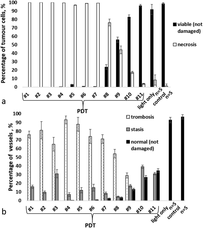
Figure 3.
Quantitative assessment of morphological changes of tumour CT-26 at 7 days post PDT: (a) percentage of viable and necrotic cells in the tumour; (b) percentage of microvascular damages in the tumour. The result are shown as mean ± SD. Notice that in case of complete (necrotic) cell death, thrombosis dominated over the other types of microvascular damages.

