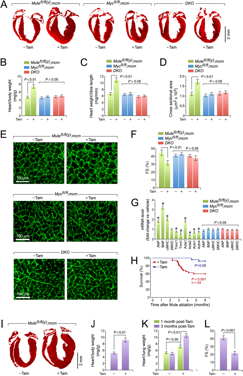
Figure 2. Genetic co-ablation of Myc and Mule prevents the cardiomyopathy associated with Mule-deficiency.
(A) Representative masson staining of longitudinal cardiac sections of the indicated mice at 4 weeks post-Tam. (B) Heart-weight corrected for body weight of the indicated strains at 4 weeks post-Tam. Animals were 15 weeks old at the time of analysis. n = 28. (C) Heart-weight corrected for tibia length at 4 weeks post-Tam. n = 28. (D) Quantification of cross-sectional area of adult cardiomyocytes at 4 weeks post-Tam. n = 12. (E) Immunofluorescence microscopy of wheat germ agglutinin (WGA; green) stained formalin-fixed LV sections at 4 weeks post-Tam. (F) Fractional shortening (FS) determined by M-mode echocardiography at 4 weeks post-Tam. n = 4. (G) Expression levels of hypertrophic and sarcomeric marker genes atrial natriuretic factor (ANP), brain natriuretic factor (BNP), α-myosin heavy chain (β-MHC), β-myosin heavy chain (β-MHC), troponin C (Tnnc1), troponin T (Tnnt1), α-actin (Acta1/2) and α-actinin (Actn1/4) as analyzed by RT-qPCR at 4 weeks post-Tam. n = 4. *P < 0.05 versus −Tam. #P < 0.01 versus −Tam. (H) Acute genetic ablation of Mule evokes premature death. Kaplan-Meier survival curves of conditional Mulefl/fl(y);mcm mice. (I) Representative masson staining of longitudinal cardiac sections from Mulefl/fl(y);mcm mice at 3 months post-Tam. (J) Heart-weight corrected for body weight of Mulefl/fl(y);mcm mice at 3 months post-Tam. n = 14. (K) Lung-weight corrected for tibia length of Mulefl/fl(y);mcm mice at the indicated time points post-Tam. n = 14. (L) Fractional shortening (FS) determined by M-mode echocardiography of Mulefl/fl(y);mcm mice at 3 months post-Tam. n = 4. Data are means ± s.e.m.

