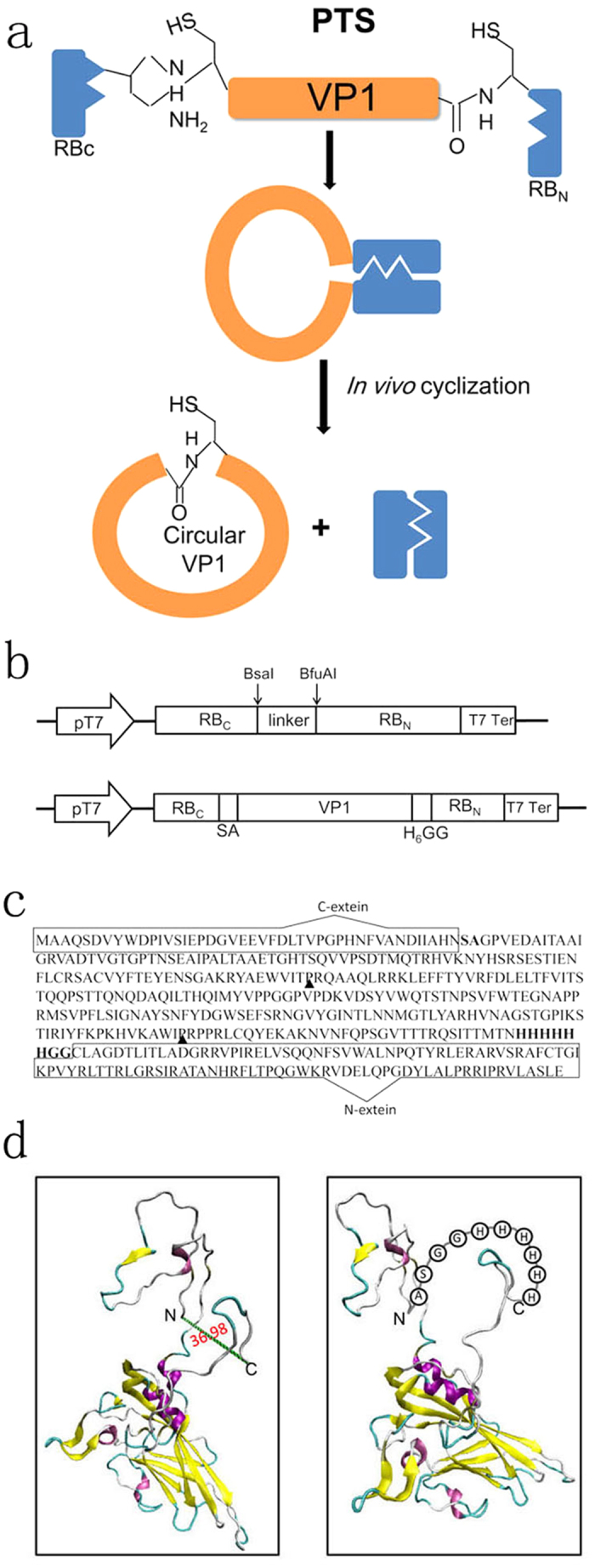
Figure 1. Protein cyclization in vivo using intramolecular trans-splicing activity of Rma DnaB intein.

(a) VP1 is sandwiched between the C-terminal fragment (RBC) and N-terminal fragment (RBN) of Rma DnaB intein. Splicing mediates the ligation of the N and C termini of VP1 through a native peptide bond. (b) Schematic representation of expression vector pERBC-RBN and pERBC-VP1-RBN. (c) Amino acid sequence of the fusion protein RBc-VP1-RBN. The C-terminal 43-residue segment (RBC) and the N-terminal 104-residue segment (RBN) of the Rma DnaB intein are enclosed with a rectangle. The linker sequence of SA and H6-GG is bold and the thrombin-specific cleavage sites marked with black arrow. (d) Models of the 3-D structure of linear (left) and circular form of VP1. The models were created based on the coordinates of the PDB code 1COV. N and C indicate N- and C-termini in the linear form.
