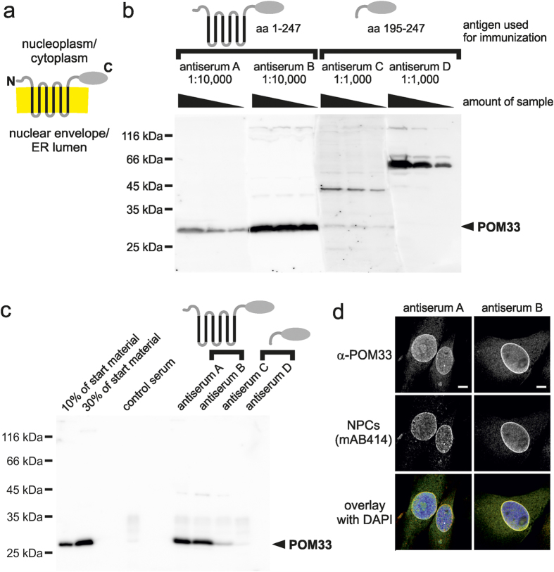
Figure 1. Antisera against full-length POM33 outperform antisera generated against a soluble fragment.
(a) Schematic representation of POM33. Predicted transmembrane regions are indicated in black, the lipid bilayer in yellow. (b) 10 μg, 3 μg and 1 μg of a total membrane fraction from Xenopus egg extracts were separated on a 12% SDS-PAGE and analyzed by western blotting using antisera against full-length POM33 (antiserum A and B) in a 1:10,000 dilution and antisera against the C-terminal domain (antiserum C and D) in a 1:1,000 dilution. Molecular size markers as well as the position of POM33 are indicated. Please note that for this analysis the blot membrane was after transfer divided into four parts, which were separately incubated with the indicated antisera. For blot analysis by ECL the parts were realigned and imaged as a whole. (c) POM33 was immunprecipitated from a solubilized total membrane fraction from Xenopus egg extracts using antisera against full-length POM33 (antiserum A and B) and antisera against the C-terminal domain (antiserum C and D). Antibody bound proteins were eluated with SDS sample buffer and separated together with 10% and 30% of the corresponding starting material on a 12% SDS-PAGE. After western blotting samples were analyzed with α-POM33 antiserum B. (d) Immunofluorescence detection of POM33. Xenopus S3 cells were fixed with 2% paraformaldehyde and stained with α-POM33 antisera A and B. Samples were co-stained with the nuclear pore complex (NPC) marker mAB414 and DAPI and analyzed by confocal microscopy. Bars: 5 μm.

