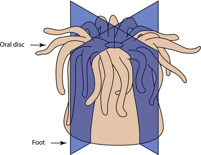
Figure 1. Diagrammatic depiction of the planes through which anemones were cut.

Blue coloured longitudinal planes indicate where each C. polypus individual (n = 3) was precisely cut from oral disc (top) to foot (bottom), resulting in four equally sized quarters with the same representation of tissues and cell types. Each fragment was sampled to represent a separate time point (0, 3, 20, and 96 hours post sectioning).
