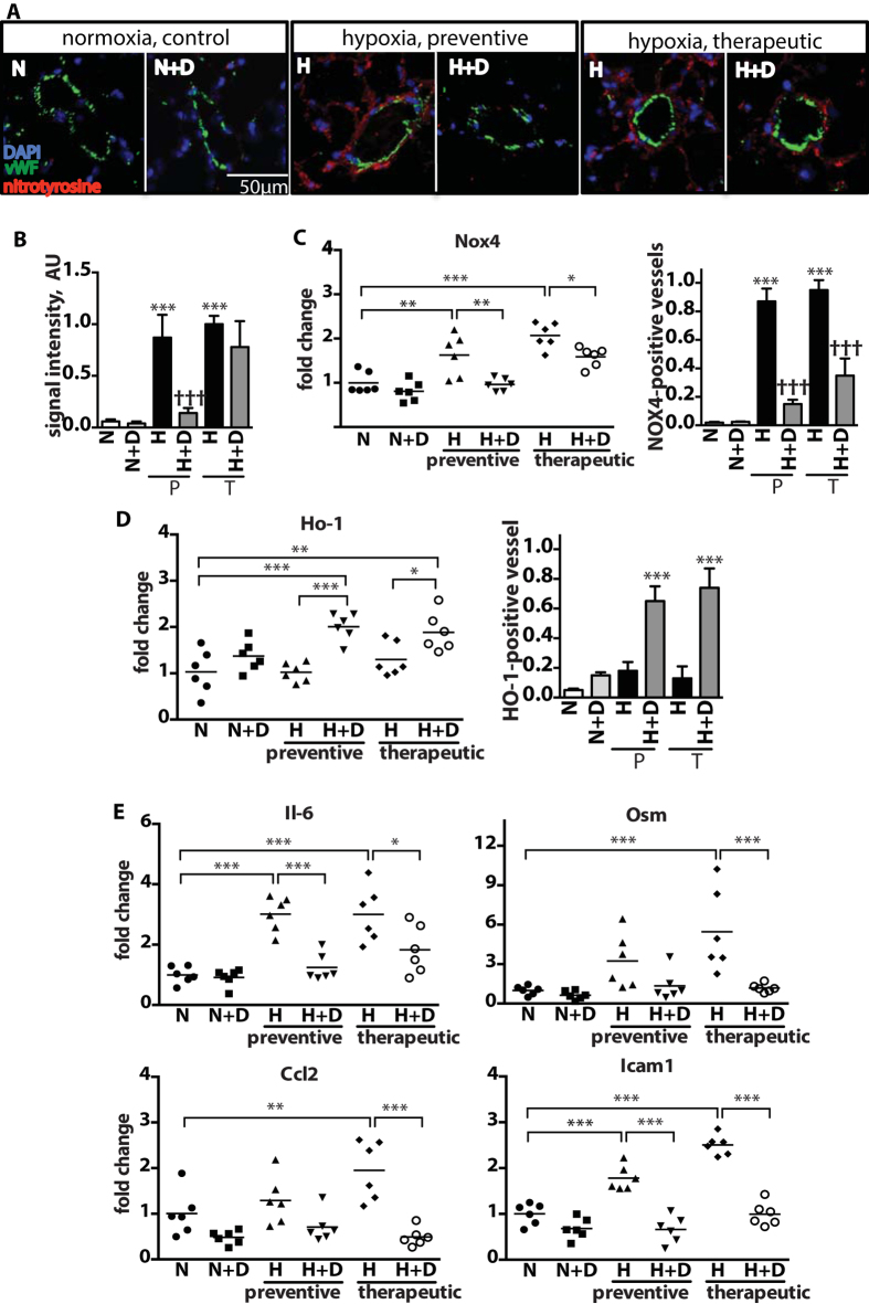
Figure 2. Attenuation of hypoxia-induced pulmonary oxidative stress damage and inflammation by DMF treatment.
(A) Representative pictures of immunofluorescent staining of mouse lungs using anti-vWF antibody and anti-nitrotyrosine antibody as a marker of oxidative damage. (B) Quantification of the mean fluorescence intensity of nitrotyrosine staining from 5 fields of view per mouse. n = 4 mice per group. normoxia (N), hypoxia (H), DMF treatment (D), preventive treatment (P), therapeutic treatment (T). (C,D,E) qPCR quantification of relative mRNA levels in a whole lung. (C) Gene expression and quantification of vessels immunoreactive for NOX4 and (D) HO-1. Vessels were counted from 5 fields of view per mouse. n = 4 mice per group. *P versus normoxia (N); †P versus hypoxia (H). (E) Gene expression of Il-6 family cytokines: Il-6 and Osm, Ccl2, and Icam1. n = 6 mice per group. Data shown as mean ± SD. *P < 0.05, **P < 0.01, ***P < 0.001.

