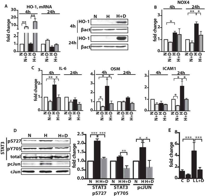
Figure 3. DMF inhibits pro-inflammatory gene expression in endothelial cells by suppressing NFκB, STAT3 and cJUN signaling.
(A,B,C,E) HPAECs were incubated for up to 24 h in (H) hypoxia (2.5% O2) or (N) normoxia (21% O2) with 10 μM DMF (D) or DMSO. Relative gene expression was measured with qPCR: (A) HO-1, (B) Nox4, (C) pro-inflammatory genes: OSM, IL-6 and ICAM1. n = 4 independent experiments. (D) 10 μM DMF treatment reduced the phosphorylation of STAT3 and cJUN in HPAECs exposed to hypoxia for 4 h. Representative immunoblots and densitometry quantification are shown. n = 3 independent experiments. (E) NFκB activity luciferase reporter assay was performed in HPAECs in presence of 10 ng/ml LPS (L) and/or 30 μM DMF (D), control cells treated with DMSO (C). n = 3 independent experiments. Data shown as mean ± SD. *P < 0.05, **P < 0.01, ***P < 0.001. The uncropped images of selected immunoblots are shown in Supplementary Fig.14.

